Royal Collection exhibition in Edinburgh will show how accurate Renaissance polymath's sketches were
Insights into the workings of the human body that Leonardo da Vinci could only obtain by dissecting scores of corpses and recording the results in exquisite drawings will be displayed for the first time beside modern 3D films, CT and MRI scans, which show how close the Renaissance genius got to the truth of what lies under the skin.
Some of the Leonardo drawings from the Royal Collection – which owns one of the greatest collections of his work in the world – have never been exhibited before in Britain, including the complete set of 18 sheets known as Anatomical Manuscript A, on which he crammed more than 240 drawings and some 13,000 words of notes in his unmistakable mirror-writing.
Leonardo was a polymath, renowned as a painter but also fascinated by science, mathematics, artillery, architecture, music, poetry, engineering and astronomy, among many other subjects. Martin Clayton, curator of the exhibition – which will open in the Queen's Gallery at Holyroodhouse in Edinburgh to coincide with the Edinburgh festival, and run on until mid-November – believes his work on anatomy could have transformed medical knowledge, but the results remained in his own notebooks until his death in 1519. He had intended to publish a major treatise but, like so much of his work, it was never completed as he was constantly distracted by new projects.
All the drawings had been imported into England by the 17th century, bound into an album which was probably bought by Charles II, and have been in the Royal Collection since at least 1690.
Some of the anatomical drawings were exhibited in the Queen's Gallery in London last year, but the Edinburgh show will be the first to compare Leonardo's results with scalpel and pen with the best results of modern technology.
Clayton said: "For the first time we will be displaying the artist's works alongside stunning examples of medical imaging, showing how the concerns and methods of the world's leading anatomists have changed little in 500 years, and how truly groundbreaking Leonardo's work was.
The exhibition will show how close Leonardo got in some of his last medical experiments to discovering the role of the beating heart in the circulation of the blood, a century before William Harvey worked it out.
In 1508 he dissected a 100-year-old man, and recorded accurately for the first time cirrhosis of the liver, and narrowing of the arteries. In the winter of 1510-11 he worked with the professor of anatomy dissecting 20 corpses in the medical school of Pavia, and covered his sheets of paper with multi-layered drawings from different angles of almost every bone in the human body including the first accurate depiction of the spine, and many of the major muscle groups.
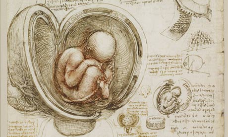
One of the more famous drawings in the collection, of a baby in the womb – partly based on his dissection of a pregnant cow – will be displayed beside a 3D ultrasound scan of a foetus.
His drawings of a hand, beginning with the bones, adding the deep muscles of the palm and then the layers of tendons, will be displayed beside a film of a dissected hand in high definition 3D. His studies of the muscles of the shoulder and arm will also be compared with an animation of the same sequence, and a 3D film of a dissected shoulder which confirms the startling accuracy he achieved.
No comments:
Post a Comment